
Embargoed till: 0300 AEDT Thu 7 Dec
Human fingers and toes do not develop outward as you would possibly count on. As a substitute, our dexterous digits are ‘sculpted’ inside a bigger foundational bud.
Now the primary human cell atlas of early limb growth has eventually revealed in beautiful element precisely how that occurs.
Previous to this, our understanding of vertebrate limb growth has been largely based mostly on mannequin organisms, resembling mice and hen embryos, and lab-grown stem cells.
Though people share some similarities with different vertebrates, their biology clearly diverges from ours.
The small print of early limb formation have additionally been rendered somewhat fuzzy by technological limitations, now surpassed, and restrictions on the use of human embryos for research beyond 14 days, a rule that has been relaxed beneath strict moral provisions.
The image constructed to this point had limbs initially emerging as shapeless limb buds protruding from the perimeters of the embryonic physique. Eight weeks later, if all goes to plan, these pouches have reworked into anatomically distinct, recognizable limbs, full with fingers and toes.
It is a exceptional course of in early embryonic growth that produces arguably considered one of our most defining human features: our long, slender, opposable thumbs.
In 2014, scientists described how particular molecules expressed at exact moments in embryonic growth moulded the formation of fingers and toes, though these predictions had been based mostly on simulations of experimental knowledge.
Now, a world workforce led by cell biologist Bao Zhang at Solar Yat-sen College in China, has coloured in that course of in beautiful element, by analyzing hundreds of single cells from donated embryonic tissues that had been between 5 and 9 weeks of growth.
“We identified 67 distinct cell clusters from 125,955 captured single cells, and spatially mapped them across four first trimester timepoints to shed new light on limb development,” the workforce writes of their printed paper.
“In doing so, we uncovered several new cell states,” they add.
“What we reveal is a highly complex and precisely regulated process,” says Hongbo Zhang, senior creator and cell biologist from Solar Yat-sen College in China.
“It is like watching a sculptor at work, chiseling away at a block of marble to reveal a masterpiece. In this case, nature is the sculptor, and the result is the incredible complexity of our fingers and toes.”
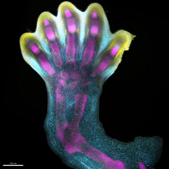
As you possibly can see within the video under, the researchers mapped gene expression patterns to see how these genetic directions formed how digits shaped.
From hazy beginnings, the expression of IRX1 (represented in aqua within the video under), a gene important for digit formation, and SOX9 (represented in magenta within the video), a gene important for skeletal growth, overlap in 5 distinct lengths inside the growing limb.
frameborder=”0″ allow=”accelerometer; autoplay; clipboard-write; encrypted-media; gyroscope; picture-in-picture; web-share” allowfullscreen>
At around 7 weeks of development, programmed cell death instructions are switched on in the undifferentiated cells congregating between these lengths (associated with the expression of MSX1, represented in yellow in the video), and well-defined fingers and toes are revealed.
Like a block of marble being sculpted into a masterpiece by the expression of these genes, our fingers and toes are chiseled out from tip to base as unneeded cells recede.
Small irregularities in this process can lead to limb deformities, which 1 in 500 people are born with – making them some of the most frequently reported syndromes at birth.
The researchers also mapped the expression of genes linked with congenital conditions, such as short fingers (brachydactyly) or webbed digits (syndactyly), to get a better sense of where limb development gets off course.
“For the primary time, we’ve got been in a position to seize the exceptional means of limb growth right down to single-cell decision in house and time,” says Sarah Teichmann, senior author and computational biologist at the Wellcome Sanger Institute.
She says creating single-cell atlases is “deepening our understanding of how anatomically advanced constructions kind, serving to us uncover the genetic and mobile processes behind wholesome human growth, with many implications for analysis and healthcare.”
Importantly, the researchers also showed that limb formation in humans and mice does follow similar trajectories, with some differences in activated genes and cell types.
The study has been published in Nature.

